This is final of "How Protect Our teeths"
Posted Under:
This is final video How Protect Our teeths. i hope this fun clip kids'll like and save their teeths.
Brush Your teeth!
Posted Under:
Brush your teeth carefully! Hey freinds this is song and helpful video to kids and childrens to protect their teeths from damages.
We Should Brush Our teeth
Posted Under:
This is topic about "We Should Brush our teeth" We must brush our teeth two times. First one is morning and second time is before every night we go to bed.
How Brush your teeth?
Posted Under:
Hello kids kutties, this is for you... how brush your theeth carefully, and protect your teeth from jems and other bacterias, I hope this willl helpful to you.
Spring Catapult
Posted Under:

The greeks continued to improve their catapult designs. In addition to simply flexing and bending a rod in order to produce force, they added more energy.
They also began to use springs made of large ropes of animal hair. The soldiers pulled back on the handle, and the "hair spring" became twisted, strong up energy. when the spring was released, the catapult shot rocks, arrows, or burning tar onto enemies.
Levers: The Product of Ancient Greek Warfare
Posted Under:
In many cases, we can't identify the inventors of ancient machines. Just as we don't know who invented the winch, we don't know the name of the Greeck soldier who invented catapult.
And yet his invention brought his people many victories in ancient battles.

The Greeks were among the first people to use catapults in warfare. They clearly inted that catapults wouldbe used to hurl an object over a long distance with great force.
Catapults are powerful and clever devices, but they're nothing more that simple leavers. To see how a catapult works, think of a screwdriver.
Have you ever used one to pry open a can of paint? The metal lid is wedged in tight, but if you use a screwdriver as a lever to pry the lid off, the can is easy to open.
Winches in Ancient China
Posted Under:
One of the most basic human needs is that for water. people living in ancient times spent a great deal of time thinking about how to gater and store water.

The need for a daily supply of water limited the number of places in which people clould live. Many groups settled near streams, rivers, and lakes.

If people wanted to live somewhere that was not close to a body of water, they had to find another way to meet their daily water needs.
Eventually, it was discovered that rivers and lakes exist underground. People began digging deep wells in order to use the groun water supply. However , pulling the water out of the wells required the development of another simple machine.
Ramps in Ancient Egypt
Posted Under:
Have you ever watched people unload their belongings from a moving van? The ramp propped onto the back of a moving van is a simple machine. A ramp is an inclined plane that works by reducing the amount of force needed to move an object.

scientists believe the ancient Egyptains were well aware of the concept of the ramp and used it to make their work easier while building the pyramids.
suppose that you are the manager of a construction team in ancient Egypt. Your team has to lift a boulder weighing 5000LB 50 ft into the air. How will you do it?
If the team tried to lift boulder straight up, they would have to apply 250,000 lb of force to it. Impossible! But suddenly, you have the idea of building a 200-ft slope leading up to the construction site, and the idea.
Machines Of the Ancient World
Posted Under:
Introduction
These days, we use many impressive technical gadgets in our everyday lives. Just think of all of these electrical devices you use; televisions, stereos, compuiters, cellulae phones- the list goes on.
You might think that the word technology applies only to electronics. However, technology, as it is discussed in science, refers to more than just Your DVD Player.
 Technology includes any tools that we use to make life easier. In fact, long before the invention of the computer, humans were clever inventors. Many of the tools we use today are based on devices that ere invented thousands of years ago.
These days, we use many impressive technical gadgets in our everyday lives. Just think of all of these electrical devices you use; televisions, stereos, compuiters, cellulae phones- the list goes on.
You might think that the word technology applies only to electronics. However, technology, as it is discussed in science, refers to more than just Your DVD Player.

Ear Anatomy pt-4
Posted Under:
This is best ear anatomy all of these video tutorials. I hope you'll like it if you want more informations. Visit here "www.myscienz.blogspot.com"
Ear anatomy pt-3 the Corti
Posted Under:
The Corti is a organ it has in middle ear. It contains auditory inner and outside hair cells. It's blog other unkown particles dusts. Save the ear.
Ear anatomy - pt2
Posted Under:
This is the Ear Anatomy pt-2 inside the ear, how indisde the organs functions you're able to watch that here and tell about this.
Ear Anatomy pt-1
Posted Under:
Hello freinds! THis is about Ear anatmoy. Our organ ear is very import to us for listen voice, noise or other sounds from vibrattions.
Was Worng about Falling Objects?
Posted Under:

Have you ever dropped a penny and a quarter at the same time from the same height? Try it, and you will see that the two coins hit the ground at the same time. This results doesnot agree with aristotle's ideas. according to aristotle, the heavier coin should strike the ground first.
Problems with Aristotle's Ideas
Posted Under:
Aristitle's ideas were accepted as the truth for hundreds of years. At a quick glance, his ideas seem to be common sense. For example, a rock dropped from an outstretched hand does fall in a straight line.
it seems logical that a heavy stone should fall faster than a much lighter block of wood. However, there are problems with aristotle's ideas.

Think about an archer who shoots a flaming arrow into the night sky.
The Curving path of the arrow's flight is shown in the illustration below. This curved path does not agree with Aristotle's belief that all motion on earth must be in a straight line.
Ideas about Weight and Falling Speed
Posted Under:

Aristotle's interest in the structure of living things was encouraged by his father. That intrest helped Aristotle develop strong observation skills. He went on to apply his observation skills to a wide range of topics.
Those topics included politics,nature,ethics,astronomy,and physics.
In time, Many of Aristotle's teachings in physics would be proved wrong. His errors included thinging that
- All motion on Earth occurs in a strainght line(called linear motion)
- an object continues in motion only as long as someting acts on it to keep it moving.
- a heavy object falls faster than a light object.
- Earth is at the center of the Universe.
Thinking About Motion
Posted Under:
Aristotle And Motion
Thin about dropping a rock while you are walking. What do you expert to happen? You expect the rock to fall and hit the ground.
But why does this happen? Is some strange force at work?

What path does the rock follow on its way to the ground? does it fall straight down? In ancient times, people known as philosophers through about questions like these.
In the says of the ancient philosophers, there were no microscopes or telescopes. No one could make exact measurements. People didn't do controlled scientific experiments. In those days, Philosophers depended on the power of the mind to find truth. The greek philosopher Aristotle was one of the most important of these early thinkers.
Aristotle was the son of a medical doctor. He was born in 384 B.C in northern Greece. He went to school at Plato's Academy. Then he became a teacher there.
Basic of Eye Anatomy
Posted Under:
This is Basic of eye anatmoy! it will useful to doctors and medical students and science students!
This is Physiology of the Eye
Posted Under:
This video simply and useful to say about physiology of the eye!
Eye Quantel Medical Anatomy
Posted Under:
Hello freinds this is about eye anatomy,Quantel Medical proposes a 3D animation explaining the anatomy and function of the eye. Vision is a fundamental and particularly complex function provided by the visual system. Main element of this system, the eye is often compared to a camera. So the eye can be described as a dark room equipped of a double objective (cornea, crystalline lens), a focus (accommodation) and a diaphragm (pupil). His photographic film (retina) which will print the image, is scanned by a cable (optic nerve) and then interpreted by the brain. Find more information about the eye
Ultrasound, Nanoparticles May Help Diabetics Avoid the Needle
Posted Under:
This world has imporved with new technologies....A new nanotechnology-based technique for regulating blood sugar in diabetics may give patients the ability to release insulin painlessly using a small ultrasound device, allowing them to go days between injections -- rather than using needles to give themselves multiple insulin injections each day. The technique was developed by researchers at North Carolina State University and the University of North Carolina at Chapel Hill

And This is hopefully a big step toward giving diabetics a more painless method of maintaining healthy blood sugar levels," says Dr. Zhen Gu, senior author of a paper on the research and an assistant professor in the joint biomedical engineering program at NC State and UNC-Chapel Hill.
The technique involves injecting biocompatible and biodegradable nanoparticles into a patient's skin. The nanoparticles are made out of poly(lactic-co-glycolic) acid (PLGA) and are filled with insulin.
Each of the PLGA nanoparticles is given either a positively charged coating made of chitosan (a biocompatible material normally found in shrimp shells), or a negatively charged coating made of alginate (a biocompatible material normally found in seaweed).
When the solution of coated nanoparticles is mixed together, the positively and negatively charged coatings are attracted to each other by electrostatic force to form a "nano-network." Once injected into the subcutaneous layer of the skin, that nano-network holds the nanoparticles together and prevents them from dispersing throughout the body.
Using the new technology developed by Gu's team, a diabetes patient doesn't have to inject a dose of insulin -- it's already there. Instead, patients can use a small, hand-held device to apply focused ultrasound waves to the site of the nano-network, painlessly releasing the insulin from its de facto reservoir into the bloodstream.
The researchers believe the technique works because the ultrasound waves excite microscopic gas bubbles in the tissue, temporarily disrupting nano-network in the subcutaneous layer of the skin. That disruption pushes the nanoparticles apart, relaxing the electrostatic force being exerted on the insulin in the reservoir. This allows the insulin to begin entering the bloodstream -- a process hastened by the effect of the ultrasound waves pushing on the insulin.
Thalamus
Posted Under:

Hey do you know? "Thalamus" is a important part of the Brain. The two thalami are located in the center of the brain, one beneath each cerebral hemisphere and next to the third ventricle.
Functionally the thalami can be thought of as relay stations for nerve impulses carrying sensory information into the brain; the thalami receive these sensory inputs as well as inputs from other parts of the brain and determine which of these signals to forward to the cerebral cortex.
Mach 1000 Shock Wave Lights Supernova Remnant
Posted Under:
What is the Mach 1000 Shock wave When a star explodes as a supernova, it shines brightly for a few weeks or months before fading away. Yet the material blasted outward from the explosion still glows hundreds or thousands of years later, forming a picturesque supernova remnant. What powers such long-lived brilliance?
 In the case of Tycho's supernova remnant, astronomers have discovered that a reverse shock wave racing inward at Mach 1000 (1000 times the speed of sound) is heating the remnant and causing it to emit X-ray light.
"We wouldn't be able to study ancient supernova remnants without a reverse shock to light them up," says Hiroya Yamaguchi, who conducted this research at the Harvard-Smithsonian Center for Astrophysics (CfA)
Modern astronomers know that the event Tycho and others observed was a Type Ia supernova, caused by the explosion of a white dwarf star. The explosion spewed elements like silicon and iron into space at speeds of more than 11 million miles per hour (5,000 km/s).
When that ejecta rammed into surrounding interstellar gas, it created a shock wave -- the equivalent of a cosmic "sonic boom." That shock wave continues to move outward today at about Mach 300. The interaction also created a violent "backwash" -- a reverse shock wave that speeds inward at Mach 1000.
"It's like the wave of brake lights that marches up a line of traffic after a fender-bender on a busy highway," explains CfA co-author Randall Smith.
Colossal New Predatory Dino Terrorized Early Tyrannosaurs
Posted Under:
Hey do you know? A new species of carnivorous dinosaur -- one of the three largest ever discovered in North America -- lived alongside and competed with small-bodied tyrannosaurs 98 million years ago.

This newly discovered species, Siats meekerorum, (pronounced see-atch) was the apex predator of its time, and kept tyrannosaurs from assuming top predator roles for millions of years.
Named after a cannibalistic man-eating monster from Ute tribal legend, Siats is a species of carcharodontosaur, a group of giant meat-eaters that includes some of the largest predatory dinosaurs ever discovered. The only other carcharodontosaur known from North America is Acrocanthosaurus, which roamed eastern North America more than 10 million years earlier. Siats is only the second carcharodontosaur ever discovered in North America; Acrocanthosaurus, discovered in 1950, was the first.
"It's been 63 years since a predator of this size has been named from North America," says Lindsay Zanno, a North Carolina State University paleontologist with a joint appointment at the North Carolina Museum of Natural Sciences, and lead author of a Nature Communications paper describing the find. "You can't imagine how thrilled we were to see the bones of this behemoth poking out of the hillside."
Zanno and colleague Peter Makovicky, from Chicago's Field Museum of Natural History, discovered the partial skeleton of the new predator in Utah's Cedar Mountain Formation in 2008. The species name acknowledges the Meeker family for its support of early career paleontologists at the Field Museum, including Zanno.
The recovered specimen belonged to an individual that would have been more than 30 feet long and weighed at least four tons. Despite its giant size, these bones are from a juvenile. Zanno and Makovicky theorize that an adult Siats might have reached the size of Acrocanthosaurus, meaning the two species vie for the second largest predator ever discovered in North America. Tyrannosaurus rex, which holds first place, came along 30 million years later and weighed in at more than twice that amount.
Although Siats and Acrocanthosaurus are both carcharodontosaurs, they belong to different sub-groups. Siats is a member of Neovenatoridae, a more slender-bodied group of carcharodontosaurs. Neovenatorids have been found in Europe, South America, China, Japan and Australia. However, this is the first time a neovenatorid has ever been found in North America.
Another Human B.anatomy
Posted Under:
This is another one of human brain anatomy this video was uploaded to education purpose only! so you can get more infos.
New Model of Human Brain
Posted Under:
Hey you can see the parts in the human brain. See and think about human brain parts and their functions as well as.
Brain Anatomy pt-2
Posted Under:
This is part-2 of brain anatomy. So You're able to watch couninously!
Brain Anatomy
Posted Under:
Hello guys! This is about human brain anatomy. It's say about completly in the parts of brain.
Does Obesity Reshape Our Sense of Taste?
Posted Under:

Obesity may alter the way we taste at the most fundamental level: by changing how our tongues react to different foods.
Compared with slimmer counterparts, the plump mice had fewer taste cells that responded to sweet stimuli. What's more, the cells that did respond to sweetness reacted relatively weakly.
The findings peel back a new layer of the mystery of how obesity alters our relationship to food.
What we see is that even at this level -- at the first step in the taste pathway -- the taste receptor cells themselves are affected by obesity," Medler said. "The obese mice have fewer taste cells that respond to sweet stimuli, and they don't respond as well."Medler said it's possible that trouble detecting sweetness may lead obese mice to eat more than their leaner counterparts to get the same payoff.
Learning more about the connection between taste, appetite and obesity is important, she said, because it could lead to new methods for encouraging healthy eating.
The Era of Neutrino Astronomy Has Begun
Posted Under:
Astrophysicists using a telescope embedded in have succeeded in a quest to detect and record the mysterious phenomena known as cosmic neutrinos -- nearly massless particles that stream to Earth at the speed of light from outside our solar system, striking the surface in a burst of energy that can be as powerful as a baseball pitcher's fastball.
 Next, they hope to build on the early success of the IceCube Neutrino Observatory to detect the source of these high-energy particles, said Physics Professor Gregory Sullivan, who led the University of Maryland's 12-person team of contributors to the IceCube Collaboration. "The era of neutrino astronomy has begun," Sullivan said as the IceCube Collaboration announced the observation of 28 very high-energy particle events that constitute the first solid evidence for astrophysical neutrinos from cosmic sources.
By studying the neutrinos that IceCube detects, scientists can learn about the nature of astrophysical phenomena occurring millions, or even billions of light years from Earth, Sullivan said.
"The sources of neutrinos, and the question of what could accelerate these particles, has been a mystery for more than 100 years. Now we have an instrument that can detect astrophysical neutrinos. It's working beautifully, and we expect it to run for another 20 years."
The collaboration's report on the first cosmic neutrino records from the IceCube Neutrino Observatory, collected from instruments embedded in one cubic kilometer of ice at the South Pole, was published Nov. 22 in the science.
Evidence of Jet in Milky Way's Black Hole
Posted Under:
Hey do you know Astronomers have long sought strong evidence that Sagittarius A* (Sgr A*), center of the milky way at the center of the Milky Way, is producing a jet of high-energy particles. Finally they have found it, in new results from NASA's Chandra X-ray Observatory and the National Science Foundation's Very Large Array (VLA) radio telescope.
 The study shows the spin axis of Sgr A* is pointing in one direction, parallel to the rotation axis of the Milky Way, which indicates to astronomers that gas and dust have migrated steadily into Sgr A* over the past 10 billion years. If the Milky Way had collided with large galaxies in the recent past and their central black holes had merged with Sgr A*, the jet could point in any direction.
The jet appears to be running into gas near Sgr A*, producing X-rays detected by Chandra and radio emission observed by the VLA. The two key pieces of evidence for the jet are a straight line of X-ray emitting gas that points toward Sgr A* and a shock front -- similar to a sonic boom -- seen in...

The jet appears to be running into gas near Sgr A*, producing X-rays detected by Chandra and radio emission observed by the VLA. The two key pieces of evidence for the jet are a straight line of X-ray emitting gas that points toward Sgr A* and a shock front -- similar to a sonic boom -- seen in...
Neanderthal Viruses Found in Modern Humans
Posted Under:

Around 8% of human DNA is made up of 'endogenous retroviruses' (ERVs), DNA sequences from viruses which pass from generation to generation. This is part of the 90% of our DNA with no known function, sometimes called 'junk' DNA.
'I wouldn't write it off as "junk" just because we don't know what it does yet,' said Dr Gkikas Magiorkinis, an MRC Fellow at Oxford University's Department of Zoology. 'Under certain circumstances, two "junk" viruses can combine to cause disease -- we've seen this many times in animals already. ERVs have been shown to cause cancer when activated by bacteria in mice with weakened immune systems.'
Dr Gkikas and colleagues are now looking to further investigate these ancient viruses, belonging to the HML2 family of viruses, for possible links with cancer and HIV.
'How HIV patients respond to HML2 is related to how fast a patient will progress to AIDS, so there is clearly a connection there,' said Dr Magiorkinis, co-author of the latest study. 'HIV patients are also at much higher risk of developing cancer, for reasons that are poorly-understood. It is possible that some of the risk factors are genetic, and may be shared with HML2. They also become reactivated in cancer and HIV infection, so might prove useful as a therapy target in the future.'
Introduction to Male Reproductive Anatomy - Part 3 - The Penis
Posted Under:
Introduction to Male Reproductive Anatomy - Part 3 - The Penis. i hope it will helpful to you 3D anatomy tutorial on the basic anatomical structure of the penis, using the BioDigital Human
Introduction to Male Reproductive Anatomy - Part 2 - Vas Deferent
Posted Under:
You can understand about this "Introduction to Male Reproductive Anatomy - Part 2 - Vas Deferen"This tutorials covers the vas (ductus) deferens, the prostate gland, the seminal vesicles, the bulbourethral glands, and the different parts of the urethra.
Introduction to Male Reproductive Anatomy - Part 1 - Testis
Posted Under:
This is about Introduction to Male Reproductive Anatomy - Part 1 - Testis , It will clear your doubts about this topic! So it will continue...
Circle of Willis - 3D Anatomy Tutorial
Posted Under:
Hey! This is Circle of Willis - 3D Anatomy Tutorial will helpful to you and medical students and doctors.
Modern Bacteria Can Add DNA from Creatures Long-Dead to Its Own
Posted Under:
From a bacteria’s perspective the environment is one big DNA waste yard. Researchers have now shown that bacteria can take up small as well as large pieces of old DNA from this scrapheap and include it in their own genome. This discovery may have major consequences – both in connection with resistance to antibiotics in hospitals and in our perception of the evolution of itself.
 Furthermore old DNA is not limited to only returning microbes to earlier states. Damaged DNA can also create new combinations of already functional sequences. You can compare it to a bunch of bacteria which poke around a trash pile looking for fragments they can use. Occasionally they hit some ‘second-hand gold’, which they can use right away. At other times they run the risk of cutting themselves up. It goes both ways. This discovery has a number of consequences partially because there is a potential risk for people when pathogen bacteria or multi-resistant bacteria exchange small fragments of ‘dangerous’ DNA e.g. at hospitals, in biological waste and in waste water.
In the grand perspective the bacteria’s uptake of short DNA represents a fundamental evolutionary process that only needs a growing cell consuming DNA pieces. A process that possibly is a kind of original type of gene-transfer or DNA-sharing between bacteria. The results show how genetic evolution can happen in jerks in small units. The meaning of this is great for our understanding of how microorganisms have exchanged genes through the history of life. The new results also support the theories about gene-transfer as a decisive factor in life’s early evolution.
Søren Overballe-Petersen explains: "This is one of the most exciting perspectives of our discovery. Computer simulations have shown that even early bacteria on Earth had the ability to share DNA – but it was hard to see how it could happen. Now we suggest how the first bacteria exchanged DNA. It is not even a mechanism developed to this specific purpose but rather as a common process, which is a consequence of living and dying."
Amber Provides New Insights Into the Evolution of Earth's Atmosphere: Low Oxygen Levels for Dinosaurs
Posted Under:

The study has been published in the journal Geochimica et Cosmochimica Acta. The interdisciplinary team, consisting of mineralogists, paleontologists and geochemists, use the preserving properties of plant resins, caused by polymerization, for their study. "During photosynthesis plants bind atmospheric carbon, whose isotopic composition is preserved in resins over millions of years, and from this, we can infer atmospheric oxygen concentrations," explains Ralf Tappert. The information about oxygen concentration comes from the isotopic composition of carbon or rather from the ratio between the stable
Why Men's Noses Are Bigger Than Women's
Posted Under:
Hello freinds do you know...Human noses come in all shapes and sizes. But one feature seems to hold true: Men's noses are bigger than women's

oxygen for muscle tissue growth and maintenance. Larger noses mean more oxygen can be breathed in and transported in the blood to supply the muscle.
It also explains why our noses are smaller than those of our ancestors, such as the Neanderthals. The reason, the researchers believe, is because our distant lineages had more muscle mass, and so needed larger noses to maintain that muscle. Modern humans have less lean muscle mass, meaning we can get away with smaller noses.
"So, in humans, the nose can become small, because our bodies have smaller oxygen requirements than we see in archaic humans," Holton says, noting also that the rib cages and lungs are smaller in modern humans, reinforcing the idea that we don't need as much oxygen to feed our frames as our ancestors. "This all tells us physiologically how modern humans have changed from their ancestors."
Holton and his team tracked nose size and growth of 38 individuals of European descent enrolled in the Iowa Facial Growth Study from three years of age until the mid-twenties, taking external and internal measurements at regular intervals for each individual. The researchers found that boys and girls have the same nose size
The Circulatory Song!
Posted Under:
Hello freinda and fans This song is really funny and we heard/watched it for the first time in science which was part of our human biology unit! If you actually learn to memorize it it can be really helpful! LOL! So hope you enjoy!
Oxygen Movement from Alveoli to Capillaries
Posted Under:
Another this one for you....Watch as a molecule of oxygen makes its way from the alveoli (gas layer) through various liquid layers in order to end up in the blood. Rishi is a pediatric infectious disease physician and works at Khan Academy. These videos do not provide medical advice and are for informational purposes only. The videos are not intended to be a substitute for professional medical advice, diagnosis or treatment. Always seek the advice of a qualified health provider with any questions you may have regarding a medical condition. Never disregard professional medical advice or delay in seeking it because of something you have read or seen in any Khan Academy video
Central Chemoreceptors
Posted Under:
Hey this is about...Central Chemoreceptors!WHat about these..Find out how the your body uses special cells that are central to the brain (inside the brain) to sense levels of CO2 and pH. Rishi is a pediatric infectious disease physician and works at Khan Academy. These videos do not provide medical advice and are for informational purposes only. The videos are not intended to be a substitute for professional medical advice, diagnosis or treatment. Always seek the advice of a qualified health provider with any questions you may have regarding a medical condition. Never disregard professional medical advice or delay in seeking it because of something you have read or seen in any Khan Academy video.
The Respiratory Center
Posted Under:
Hello freinds! Find out how the respiratory center collects information from all over the body and then helps regulate your breathing. Rishi is a pediatric infectious disease physician and works at Khan Academy. These videos do not provide medical advice and are for informational purposes only. The videos are not intended to be a substitute for professional medical advice, diagnosis or treatment. Always seek the advice of a qualified health provider with any questions you may have regarding a medical condition. Never disregard professional medical advice or delay in seeking it because of something you have read or seen in any Khan Academy video
New Insights Into Brain Neuronal Networks
Posted Under:

Using brain-wide and consistent tracer data, the researchers describe the cortex as a network of connections with a "bow tie" structure characterized by a high-efficiency, dense core connecting with "wings" of feed-forward and feedback pathways to the rest of the cortex (periphery). The local circuits, reaching to within 2.5 millimeters and taking up more than 70 percent of all the connections in the macaque cortex, are integrated across areas with different functional modalities (somatosensory, motor, cognitive) with medium- to long-range projections.
The authors also report on a simple network model that incorporates the physical principle of entropic cost to long wiring and the spatial positioning of the functional areas in the cortex. They show that this model reproduces the properties of the connectivity data in the experiments, including the structure of the bow tie. The wings of the bow tie emerge from the counterstream organization of the feed-forward and feedback nature of the pathways. They also demonstrate that, contrary to previous beliefs, such high-density cortical graphs can achieve simultaneously strong connectivity (almost direct between any two areas), communication efficiency, and economy of connections (shown via optimizing total wire cost) via weight-distance correlations that are also consequences of this simple network model.
This bow tie arrangement is a typical feature of self-organizing information processing systems. The paper notes that the cortex has some analogies with information-processing networks such as the World Wide Web, as well as metabolism, the immune system and cell signaling. The core-periphery bow tie structure, they say, is "an evolutionarily favored structure for a wide variety of complex networks" because "these systems are not in thermodynamic equilibrium and are required to maintain energy and matter flow through the system." The brain, however, also shows important differences from such systems. For example, destination addresses are encoded in information packets sent along the Internet, apparently unlike in the brain, and location and timing of activity are critical factors of information processing in the brain, unlike in the Internet.
"Biological data is extremely complex and diverse," Toroczkai said. "However, as a physicist, I am interested in what is common or invariant in the data, because it may reveal a fundamental organizational principle behind a complex system. A minimal theory that incorporates such principle should reproduce the observations, if not in great detail, but in extent. I believe that with additional consistent data, as those obtained by the Kennedy team, the fundamental principles of massive information processing in brain neuronal networks are within reach."
Is Global Heating Hiding out in the Oceans? Parts of Pacific Warming 15 Times Faster Than in Past 10,000 Years
Posted Under:

"We're experimenting by putting all this heat in the ocean without quite knowing how it's going to come back out and affect climate," said study coauthor Braddock Linsley, a climate scientist at Columbia University's Lamont-Doherty Earth Observatory. "It's not so much the magnitude of the change, but the rate of change."
In its latest report, released in September, the UN's Intergovernmental Panel on Climate Change (IPCC) noted the recent slowdown in the rate of global warming. While global temperatures rose by about one-fifth of a degree Fahrenheit per decade from the 1950s through 1990s, warming slowed to just half that rate after the record hot year of 1998. The IPCC has attributed the pause to natural climate fluctuations caused by volcanic eruptions, changes in solar intensity, and the movement of heat through the ocean. Many scientists note that 1998 was an exceptionally hot year even by modern standards, and so any average rise using it as a starting point would downplay the longer-term warming trend.
The IPCC scientists agree that much of the heat that humans have put into the atmosphere since the 1970s through greenhouse gas emissions probably has been absorbed by the ocean. However, the findings in Science put this idea into a long-term context, and suggest that the oceans may be storing even more of the effects of human emissions than scientists have so far realized. "We may have underestimated the efficiency of the oceans as a storehouse for heat and energy," said study lead author, Yair Rosenthal, a climate scientist at Rutgers University. "It may buy us some time -- how much time, I don't really know. But it's not going to stop climate change."
Ocean heat is typically measured from buoys dispersed throughout the ocean, and with instruments lowered from ships, with reliable records at least in some places going back to the 1960s. To look back farther in time, scientists have developed ways to analyze the chemistry of ancient marine life to reconstruct the climates in which they lived. In a 2003 expedition to Indonesia, the researchers collected cores of sediment from the seas where water from the Pacific flows into the Indian Ocean. By measuring the levels of magnesium to calcium in the shells of Hyalinea balthica, a one-celled organism buried in those sediments, the researchers estimated the temperature of the middle-depth waters where H. Balthica lived, from about 1,500 to 3,000 feet down. The temperature record there reflects middle-depth temperatures throughout the western Pacific, the researchers say, since the waters around Indonesia originate from the mid-depths of the North and South Pacific.
Though the climate of the last 10,000 years has been thought to be relatively stable, the researchers found that the Pacific intermediate depths have generally been cooling during that time, though with various ups and downs. From about 7,000 years ago until the start of the Medieval Warm Period in northern Europe, at about 1100, the water cooled gradually, by almost 1 degree C, or almost 2 degrees F. The rate of cooling then picked up during the so-called Little Ice Age that followed, dropping another 1 degree C, or 2 degrees F, until about 1600. The authors attribute the cooling from 7,000 years ago until the Medieval Warm Period to changes in Earth's orientation toward the sun, which affected how much sunlight fell on both poles. In 1600 or so, temperatures started gradually going back up. Then, over the last 60 years, water column temperatures, averaged from the surface to 2,200 feet, increased 0.18 degrees C, or .32 degrees F. That might seem small in the scheme of things, but it's a rate of warming 15 times faster than at any period in the last 10,000 years, said Linsley.
One explanation for the recent slowdown in global warming is that a prolonged La Niña-like cooling of eastern Pacific surface waters has helped to offset the global rise in temperatures from greenhouse gases. In a study in the journal Nature in August, climate modelers at the Scripps Institution of Oceanography showed that La Niña cooling in the Pacific seemed to suppress global average temperatures during northern hemisphere winters but allowed temperatures to rise during northern hemisphere summers, explaining last year's record U.S. heat wave and the ongoing loss of Arctic sea ice.
When the La Niña cycle switches, and the Pacific reverts to a warmer than usual El Niño phase, global temperatures may likely shoot up again, along with the rate of warming. "With global warming you don't see a gradual warming from one year to the next," said Kevin Trenberth, a climate scientist at the National Center for Atmospheric Research in Boulder, Colo., who was not involved in the research. "It's more like a staircase. You trot along with nothing much happening for 10 years and then suddenly you have a jump and things never go back to the previous level again."
The study's long-term perspective suggests that the recent pause in global warming may just reflect random variations in heat going between atmosphere and ocean, with little long-term importance, says Drew Shindell, a climate scientist with joint appointments at Columbia's Earth Institute and the NASA Goddard Institute for Space Studies, and a lead author on the latest IPCC report. "Surface temperature is only one indicator of climate change," he said. "Looking at the total energy stored by the climate system or multiple indicators--glacier melting, water vapor in the atmosphere, snow cover, and so on -- may be more useful than looking at surface temperature alone."
Surprising Variation Among Genomes of Individual Neurons from Same Brain
Posted Under:
Hey It was once thought that each cell in a person's body possesses the same DNA code and that the particular way the genome is read imparts cell function and defines the individual. For many cell types in our bodies, however, that is an oversimplification. Studies of neuronal genomes published in the past decade have turned up extra or missing chromosomes, or pieces of DNA that can copy and paste themselves throughout the genomes.
 The only way to know for sure that neurons from the same person harbor unique DNA is by profiling the genomes of single cells instead of bulk cell populations, the latter of which produce an average. Now, using single-cell sequencing, Salk Institute researchers and their collaborators have shown that the genomic structures of individual neurons differ from each other even more than expected. The findings were published November 1 in Science.
"Contrary to what we once thought, the genetic makeup of neurons in the brain aren't identical, but are made up of a patchwork of DNA," says corresponding author Fred Gage, Salk's Vi and John Adler Chair for Research on Age-Related Neurodegenerative Disease.
In the study, led by Mike McConnell, a former junior fellow in the Crick-Jacobs Center for Theoretical and Computational Biology at the Salk, researchers isolated about 100 neurons from three people posthumously. The scientists took a high-level view of the entire genome -- -- looking for large deletions and duplications of DNA called copy number variations or CNVs -- -- and found that as many as 41 percent of neurons had at least one unique, massive CNV that arose spontaneously, meaning it wasn't passed down from a parent. The CNVs are spread throughout the genome, the team found.
The miniscule amount of DNA in a single cell has to be chemically amplified many times before it can be sequenced. This process is technically challenging, so the team spent a year ruling out potential sources of error in the process.
"A good bit of our study was doing control experiments to show that this is not an artifact," says Gage. "We had to do that because this was such a surprise -- -- finding out that individual neurons in your brain have different DNA content."
The group found a similar amount of variability in CNVs within individual neurons derived from the skin cells of three healthy people. Scientists routinely use such induced pluripotent stem cells (iPSCs) to study living neurons in a culture dish. Because iPSCs are derived from single skin cells, one might expect their genomes to be the same.
"The surprising thing is that they're not," says Gage. "There are quite a few unique deletions and amplifications in the genomes of neurons derived from one iPSC line."
Interestingly, the skin cells themselves are genetically different, though not nearly as much as the neurons. This finding, along with the fact that the neurons had unique CNVs, suggests that the genetic changes occur later in development and are not inherited from parents or passed to offspring.
It makes sense that neurons have more diverse genomes than skin cells do, says McConnell, who is now an assistant professor of biochemistry and molecular genetics at the University of Virginia School of Medicine in Charlottesville. "The thing about neurons is that, unlike skin cells, they don't turn over, and they interact with each other," he says. "They form these big complex circuits, where one cell that has CNVs that make it different can potentially have network-wide influence in a brain."
Spontaneously occurring CNVs have also been linked to risk for brain disorders such as schizophrenia and autism, but those studies usually pool many blood cells. As a result, the CNVs uncovered in those studies affect many if not all cells, which suggests that they arise early in development.
The purpose of CNVs in the healthy brain is still unclear, but researchers have some ideas. The modifications might help people adapt to new surroundings encountered over a lifetime, or they might help us survive a massive viral infection. The scientists are working out ways to alter genomic variability in iPSC-derived neurons and challenge them in specific ways in the culture dish.
Cells with different genomes probably produce unique RNA and then proteins. However, for now, only one sequencing technology can be applied to a single cell.
"If and when more than one method can be applied to a cell, we will be able to see whether cells with different genomes have different transcriptomes (the collection of all the RNA in a cell) in predictable ways," says McConnell.
In addition, it will be necessary to sequence many more cells, and in particular, more cell types, notes corresponding author Ira Hall, an associate professor of biochemistry and molecular genetics at the University of Virginia. "There's a lot more work to do to really understand to what level we think the things we've found are neuron-specific or associated with different parameters like age or genotype," he says.
Other authors on the study are Michael Lindberg and Svetlana Shumilina of the Department of Biochemistry and Molecular Genetics at the University of Virginia School of Medicine; Kristen Brennand, now at the Icahn School of Medicine at Mount Sinai in New York; Julia Piper, now at Harvard University in Cambridge, Massachusetts; Thierry Voet and Joris Vermeesch of the Center for Human Genetics, KU Leuven, Leuven, Belgium; Chris Cowing-Zitron of Salk's Laboratory of Genetics; and Roger Lasken of the J. Craig Venter Institute in San Diego.
This work was supported by the Crick-Jacobs Center for Theoretical and Computational Biology, the G. Harold & Leila Y. Mathers Foundation, the National Institutes of Health, the Leona M. and Harry B. Helmsley Charitable Trust, the JPB Foundation, and the Burroughs Wellcome Fund.
Fossil of Largest Known Platypus Discovered in Australia
Posted Under:

I say about this the new platypus species, named Obdurodon tharalkooschild, is based on a single tooth from the famous Riversleigh World Heritage Area of northwest Queensland. While many of Riversleigh's fossil deposits are now being radiometrically dated, the precise age of the particular deposit that produced this giant platypus is in doubt but is likely to be between 15 and 5 million years old.
"Monotremes (platypuses and echidnas) are the last remnant of an ancient radiation of mammals unique to the southern continents. A new platypus species, even one that is highly incomplete, is a very important aid in developing understanding about these fascinating mammals," said PhD candidate Rebecca Pian, lead author of the study.
Based on the size of tooth, it is estimated that this extinct species would have been nearly a meter (more than three feet) long, twice the size of the modern platypus. The bumps and ridges on the teeth also provide clues about what this species likely ate.
"Like other platypuses, it was probably a mostly aquatic mammal, and would have lived in and around the freshwater pools in the forests that covered the Riversleigh area millions of years ago," said Dr. Suzanne Hand of the University of New South Wales, a co-author of the study. "Obdurodon tharalkooschild was a very large platypus with well-developed teeth, and we think it probably fed not only on crayfish and other freshwater crustaceans, but also on small vertebrates including the lungfish, frogs, and small turtles that are preserved with it in the Two Tree Site fossil deposit."
The oldest platypus fossils come from 61 million-year-old rocks in southern South America. Younger platypus fossils are known from Australia in what is now the Simpson Desert. Before the discovery of Obdurodon tharalkooschild, these fossils suggested that platypuses became smaller and reduced the size of their teeth through time. The modern platypus completely lacks teeth as an adult and instead bears horny pads in its mouth. The name Obdurodon comes from the Greek for "lasting (obdurate) tooth" and was coined to distinguish extinct toothed platypuses from the essentially toothless modern species.
"Discovery of this new species was a shock to us because prior to this, the fossil record suggested that the evolutionary tree of platypuses was relatively linear one," said Dr. Michael Archer of the University of New South Wales, a co-author of the study. "Now we realize that there were unanticipated side branches on this tree, some of which became gigantic."
The specific epithet of the new species, tharalkooschild, honors an Indigenous Australian creation story about the origin of the platypus. In the Dreamtime, Tharalkoo was a head-strong girl duck inclined to disobey her parents. Her parents warned her not to swim downriver because Bigoon the Water-rat would have his wicked way with her. Scoffing, she disobeyed her parents and was ravished by Bigoon. By the time Tharalkoo escaped and returned to her family, the other girl ducks were laying eggs, so she did the same. But instead of a fluffy little duckling emerging from her egg, her child was an amazing chimera that had the bill, webbed hind feet, and egg-laying habit of a duck, along with the fur and front feet of a rodent -- the first Platypus.
Respiratory system with differnt clips
Posted Under:
Hello freinds hes right, the left lung has one less branch. if you notice the right lung has 3 lobes, the left has 2 lobes, and that is due to the missing or absence of one left branch on the left. also, because the heart takes up some space (cardiac notch) where the 3rd lobe would of been
Cellular Respiration
Posted Under:
Verny Lopez Nieto explains the respiratory chain in aerobic conditions in the inner mitochondrial membrane. It argues that the process of cellular respiration involves the formation of energy from ATP-APP molecules and explains the formation of NADH and FADH molecules, which are those that carry high-energy electrons. Finally, it explains the formation of potential energy.
Respiratory system
Posted Under:
Hello freinds and fans this is for you It's pretty good explanation, but is clearly not mixed with scientific information. Science studies nature, but it does not make interpretations. Evolution or not does not matter as long evolution to gain a knowledge of nature, but this is religion and no argument given here demonstrates a thorough understanding of the theory of evolution.
Lung Anatomy
Posted Under:
is 3D medical animation begins with a detailed description of the anatomy and physiology of the lungs (Pulmonary system). It describes the pleura and diaphragm which aid in lung expansion. The animation also deals with lung cancer and the role of lymph in transporting bacteria, allergens and cancer cells away from the lungs and to the lymph nodes.
Problems With Alzheimer’s Protein Can Jam Up Traffic in the Brain
Posted Under:

It has to do with how materials travel up and down brain cells, which are also called neurons.
In an Oct. 8 paper in Human Molecular Genetics, University at Buffalo researchers report that presenilin works with an enzyme called GSK-3ß to control how fast materials -- like proteins needed for cell survival -- move through the cells.
"If you have too much presenilin or too little, it disrupts the activity of GSK-3ß, and the transport of cargo along neurons becomes uncoordinated," says lead researcher Shermali Gunawardena, PhD, an assistant professor of biological sciences at UB. "This can lead to dangerous blockages."
More than 150 mutations of presenilin have been found in Alzheimer's patients, and scientists have previously shown that the protein, when defective, can cause neuronal blockages by snipping another protein into pieces that accumulate in brain cells.
But this well-known mechanism isn't the only way presenilin fuels disease, as Gunawardena's new study shows.
"Our work elucidates how problems with presenilin could contribute to early problems observed in Alzheimer's disease," she says. "It highlights a potential pathway for early intervention through drugs -- prior to neuronal loss and clinical manifestations of disease."
The study suggests that presenilin activates GSK-3ß. This is an important finding because the enzyme helps control the speed at which tiny, organic bubbles called vesicles ferry cargo along neuronal highways. (You can think of vesicles as trucks, each powered by little molecular motors called dyneins and kinesins.)
When researchers lowered the amount of presenilin in the neurons of fruit fly larvae, less GSK-3ß became activated and vesicles began speeding along cells in an uncontrolled manner.
Decreasing levels of both presenilin and GSK-3ß at once made things worse, resulting in "traffic jams" as the bubbles got stuck in neurons.
"Both GSK-3ß and presenilin have been shown to be involved in Alzheimer's disease, but how they are involved has not always been clear," Gunawardena says. "Our research provides new insight into this question."
Gunawardena proposes that GSK-3ß -- short for glycogen synthase kinase-3beta -- acts as an "on switch" for dynein and kynesin motors, telling them when to latch onto vesicles.
Dyneins carry vesicles toward the cell nucleus, while kinesins move in the other direction, toward the periphery of the cell. When all is well and GSK-3ß levels are normal, both types of motors bind to vesicles in carefully calibrated numbers, resulting in smooth traffic flow along neurons.
That's why it's so dangerous when GSK-3ß levels are off-kilter, she says.
When GSK-3ß levels are high, too many motors attach to the vesicles, leading to slow movement as motor activity loses coordination. Low GSK-3ß levels appear to have the opposite effect, causing fast, uncontrolled movement as too few motors latch onto vesicles.
Nurturing May Protect Kids from Brain Changes Linked to Poverty!
Posted Under:
Growing up in poverty can have long-lasting, negative consequences for a child. But for poor children raised by parents who lack nurturing skills, the effects may be particularly worrisome, according to a new study at Washington University School of Medicine in St. Louis.
 Among children living in poverty, the researchers identified changes in the brain that can lead to lifelong problems like depression, learning difficulties and limitations in the ability to cope with stress. The study showed that the extent of those changes was influenced strongly by whether parents were nurturing.
The good news, according to the researchers, is that a nurturing home life may offset some of the negative changes in brain anatomy among poor children. And the findings suggest that teaching nurturing skills to parents -- particularly those living in poverty -- may provide a lifetime benefit for their children.
The study is published online Oct. 28 and will appear in the November issue of JAMA Pediatrics.
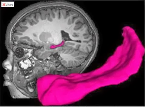
Using magnetic resonance imaging (MRI), the researchers found that poor children with parents who were not very nurturing were likely to have less gray and white matter in the brain. Gray matter is closely linked to intelligence, while white matter often is linked to the brain's ability to transmit signals between various cells and structures.
The MRI scans also revealed that two key brain structures were smaller in children who were living in poverty: the amygdala, a key structure in emotional health, and the hippocampus, an area of the brain that is critical to learning and memory.
"We've known for many years from behavioral studies that exposure to poverty is one of the most powerful predictors of poor developmental outcomes for children," said principal investigator Joan L. Luby, MD, a Washington University child psychiatrist at St. Louis Children's Hospital. "A growing number of neuroscience and brain-imaging studies recently have shown that poverty also has a negative effect on brain development.
"What's new is that our research shows the effects of poverty on the developing brain, particularly in the hippocampus, are strongly influenced by parenting and life stresses that the children experience."
Luby, a professor of psychiatry and director of the university's Early Emotional Development Program, is in the midst of a long-term study of childhood depression. As part of the Preschool Depression Study, she has been following 305 healthy and depressed kids since they were in preschool. As the children have grown, they also have received MRI scans that track brain development.
"We actually stumbled upon this finding," she said. "Initially, we thought we would have to control for the effects of poverty, but as we attempted to control for it, we realized that poverty was really driving some of the outcomes of interest, and that caused us to change our focus to poverty, which was not the initial aim of this study."
In the new study, Luby's team looked at scans from 145 children enrolled in the depression study. Some were depressed, others healthy, and others had been diagnosed with different psychiatric disorders such as ADHD (attention-deficit hyperactivity disorder). As she studied these children, Luby said it became clear that poverty and stressful life events, which often go hand in hand, were affecting brain development.
The researchers measured poverty using what's called an income-to-needs ratio, which takes a family's size and annual income into account. The current federal poverty level is $23,550 for a family of four.
Although the investigators found that poverty had a powerful impact on gray matter, white matter, hippocampal and amygdala volumes, they found that the main driver of changes among poor children in the volume of the hippocampus was not lack of money but the extent to which poor parents nurture their children. The hippocampus is a key brain region of interest in studying the risk for impairments.
Luby's team rated nurturing using observations made by the researchers -- who were unaware of characteristics such as income level or whether a child had a psychiatric diagnosis -- when the children came to the clinic for an appointment. And on one of the clinic visits, the researchers rated parental nurturing using a test of the child's impatience and of a parent's patience with that child.
While waiting to see a health professional, a child was given a gift-wrapped package, and that child's parent or caregiver was given paperwork to fill out. The child, meanwhile, was told that s/he could not open the package until the caregiver completed the paperwork, a task that researchers estimated would take about 10 minutes.
Luby's team found that parents living in poverty appeared more stressed and less able to nurture their children during that exercise. In cases where poor parents were rated as good nurturers, the children were less likely to exhibit the same anatomical changes in the brain as poor children with less nurturing parents.
"Parents can be less emotionally responsive for a whole host of reasons," Luby said. "They may work two jobs or regularly find themselves trying to scrounge together money for food. Perhaps they live in an unsafe environment. They may be facing many stresses, and some don't have the capacity to invest in supportive parenting as much as parents who don't have to live in the midst of those adverse circumstances."
The researchers also found that poorer children were more likely to experience stressful life events, which can influence brain development. Anything from moving to a new house to changing schools to having parents who fight regularly to the death of a loved one is considered a stressful life event.
Luby believes this study could provide policymakers with at least a partial answer to the question of what it is about poverty that can be so detrimental to a child's long-term developmental outcome. Because it appears that a nurturing parent or caregiver may prevent some of the changes in brain anatomy that this study identified, Luby said it is vital that society invest in public health prevention programs that target parental nurturing skills. She suggested that a key next step would be to determine if there are sensitive developmental periods when interventions with parents might have the most powerful impact.
"Children who experience positive caregiver support don't necessarily experience the developmental, cognitive and emotional problems that can affect children who don't receive as much nurturing, and that is tremendously important," Luby said. "This study gives us a feasible, tangible target with the suggestion that early interventions that focus on parenting may provide a tremendous payoff."
The good news, according to the researchers, is that a nurturing home life may offset some of the negative changes in brain anatomy among poor children. And the findings suggest that teaching nurturing skills to parents -- particularly those living in poverty -- may provide a lifetime benefit for their children.
The study is published online Oct. 28 and will appear in the November issue of JAMA Pediatrics.
Using magnetic resonance imaging (MRI), the researchers found that poor children with parents who were not very nurturing were likely to have less gray and white matter in the brain. Gray matter is closely linked to intelligence, while white matter often is linked to the brain's ability to transmit signals between various cells and structures.
The MRI scans also revealed that two key brain structures were smaller in children who were living in poverty: the amygdala, a key structure in emotional health, and the hippocampus, an area of the brain that is critical to learning and memory.
"We've known for many years from behavioral studies that exposure to poverty is one of the most powerful predictors of poor developmental outcomes for children," said principal investigator Joan L. Luby, MD, a Washington University child psychiatrist at St. Louis Children's Hospital. "A growing number of neuroscience and brain-imaging studies recently have shown that poverty also has a negative effect on brain development.
"What's new is that our research shows the effects of poverty on the developing brain, particularly in the hippocampus, are strongly influenced by parenting and life stresses that the children experience."
Luby, a professor of psychiatry and director of the university's Early Emotional Development Program, is in the midst of a long-term study of childhood depression. As part of the Preschool Depression Study, she has been following 305 healthy and depressed kids since they were in preschool. As the children have grown, they also have received MRI scans that track brain development.
"We actually stumbled upon this finding," she said. "Initially, we thought we would have to control for the effects of poverty, but as we attempted to control for it, we realized that poverty was really driving some of the outcomes of interest, and that caused us to change our focus to poverty, which was not the initial aim of this study."
In the new study, Luby's team looked at scans from 145 children enrolled in the depression study. Some were depressed, others healthy, and others had been diagnosed with different psychiatric disorders such as ADHD (attention-deficit hyperactivity disorder). As she studied these children, Luby said it became clear that poverty and stressful life events, which often go hand in hand, were affecting brain development.
The researchers measured poverty using what's called an income-to-needs ratio, which takes a family's size and annual income into account. The current federal poverty level is $23,550 for a family of four.
Although the investigators found that poverty had a powerful impact on gray matter, white matter, hippocampal and amygdala volumes, they found that the main driver of changes among poor children in the volume of the hippocampus was not lack of money but the extent to which poor parents nurture their children. The hippocampus is a key brain region of interest in studying the risk for impairments.
Luby's team rated nurturing using observations made by the researchers -- who were unaware of characteristics such as income level or whether a child had a psychiatric diagnosis -- when the children came to the clinic for an appointment. And on one of the clinic visits, the researchers rated parental nurturing using a test of the child's impatience and of a parent's patience with that child.
While waiting to see a health professional, a child was given a gift-wrapped package, and that child's parent or caregiver was given paperwork to fill out. The child, meanwhile, was told that s/he could not open the package until the caregiver completed the paperwork, a task that researchers estimated would take about 10 minutes.
Luby's team found that parents living in poverty appeared more stressed and less able to nurture their children during that exercise. In cases where poor parents were rated as good nurturers, the children were less likely to exhibit the same anatomical changes in the brain as poor children with less nurturing parents.
"Parents can be less emotionally responsive for a whole host of reasons," Luby said. "They may work two jobs or regularly find themselves trying to scrounge together money for food. Perhaps they live in an unsafe environment. They may be facing many stresses, and some don't have the capacity to invest in supportive parenting as much as parents who don't have to live in the midst of those adverse circumstances."
The researchers also found that poorer children were more likely to experience stressful life events, which can influence brain development. Anything from moving to a new house to changing schools to having parents who fight regularly to the death of a loved one is considered a stressful life event.
Luby believes this study could provide policymakers with at least a partial answer to the question of what it is about poverty that can be so detrimental to a child's long-term developmental outcome. Because it appears that a nurturing parent or caregiver may prevent some of the changes in brain anatomy that this study identified, Luby said it is vital that society invest in public health prevention programs that target parental nurturing skills. She suggested that a key next step would be to determine if there are sensitive developmental periods when interventions with parents might have the most powerful impact.
"Children who experience positive caregiver support don't necessarily experience the developmental, cognitive and emotional problems that can affect children who don't receive as much nurturing, and that is tremendously important," Luby said. "This study gives us a feasible, tangible target with the suggestion that early interventions that focus on parenting may provide a tremendous payoff."
Smart Neurons: Single Neuronal Dendrites Can Perform Computations
Posted Under:

The study, published today (Sunday) in Nature and carried out by researchers based at the Wolfson Institute for Biomedical Research at UCL, the MRC Laboratory for Molecular Biology in Cambridge and the University of North Carolina at Chapel Hill, examined neurons in areas of the mouse brain which are responsible for processing visual input from the eyes. The scientists achieved an important breakthrough: they succeeded in making incredibly challenging electrical and optical recordings directly from the tiny dendrites of neurons in the intact brain while the brain was processing visual information.
These recordings revealed that visual stimulation produces specific electrical signals in the dendrites -- bursts of spikes -- which are tuned to the properties of the visual stimulus.
The results challenge the widely held view that this kind of computation is achieved only by large numbers of neurons working together, and demonstrate how the basic components of the brain are exceptionally powerful computing devices in their own right.
Senior author Professor Michael Hausser commented: "This work shows that dendrites, long thought to simply 'funnel' incoming signals towards the soma, instead play a key role in sorting and interpreting the enormous barrage of inputs received by the neuron. Dendrites thus act as miniature computing devices for detecting and amplifying specific types of input.
"This new property of dendrites adds an important new element to the "toolkit" for computation in the brain. This kind of dendritic processing is likely to be widespread across many brain areas and indeed many different animal species, including humans."
Funding for this study was provided by the Gatsby Charitable Foundation, the Wellcome Trust, and the European Research Council, as well as the Human Frontier Science Program, the Klingenstein Foundation, Helen Lyng White, the Royal Society, and the Medical Research Council.
Need Different Types of Tissue? Just Print Them!
Posted Under:

The recent organ transplant scandals have only made the problem worse. According to the German Organ Transplantation Foundation (DSO), the number of organ donors in the first half of 2013 has declined more than 18 percent in comparison to the same period the previous year. At the same time, one can assume that the demand in the next years will continuously rise, because we continue to age and field of transplantation medicine is continuously advancing. Many critical illnesses can already be successfully treated today by replacing cells, tissue, or organs. Government, industry, and the research establishment have therefore been working hard for some time to improve methods and procedures for artificially producing tissue. This is how the gap in supply is supposed to be closed.
Bio-ink made from living cells
One technology might assume a decisive role in this effort, one that we are all familiar with from the office, and that most of us would certainly not immediately connect with the production of artificial tissue: the inkjet printer. Scientists of the Fraunhofer Institute for Interfacial Engineering and Biotechnology (IGB) in Stuttgart have succeeded in deve- loping suitable bio-inks for this printing technology. The transparent liquids consist of components from the natural tissue matrix and living cells. The substance is based on a well known biological material: gelatin. Gelatin is derived from collagen, the main constituent of native tissue. The researchers have chemically modified the gelling behavior of the gelatin to adapt the biological molecules for printing. Instead of gelling like unmodified gelatin, the bio-inks remain fluid during printing. Only after they are irradiated with UV light, they crosslink and cure to form hydrogels. These are polymers containing a huge amount of water (just like native tissue), but which are stable in aqueous environments and when being warmed up to physiological 37°C. The researchers can control the chemical modification of the biological molecules so that the resulting gels have differing strengths and swelling characteristics. The properties of natural tissue can therefore be imitated -- from solid cartilage to soft adipose tissue.
In Stuttgart synthetic raw materials are printed as well that can serve as substitutes for the extracellular matrix. For example a system that cures to a hydrogel devoid of by-products, and can be immediately populated with genuine cells. "We are concentrating at the moment on the 'natural' variant. That way we remain very close to the original material. Even if the potential for synthetic hydrogels is big, we still need to learn a fair amount about the interactions between the artificial substances and cells or natural tissue. Our biomolecule-based variants provide the cells with a natural environment instead, and therefore can promote the self-organizing behavior of the printed cells to form a functional tissue model," explains Dr. Kirsten Borchers in describing the approach at IGB.
The printers at the labs in Stuttgart have a lot in common with conventional office printers: the ink reservoirs and jets are all the same. The differences are discovered only under close inspection. For example, the heater on the ink container with which the right temperature of the bio-inks is set. The number of jets and tanks is smaller than in the office counterpart as well. "We would like to increase the number of these in cooperation with industry and other Fraunhofer Institutes in order to simultaneously print using various inks with different cells and matrices. This way we can come closer to replicating complex structures and different types of tissue," says Borchers.
The big challenge at the moment is to produce vascularized tissue. This means tissue that has its own system of blood vessels through which the tissue can be provided with nutrients. IGB is working on this jointly with other partners under Project ArtiVasc 3D, supported by the European Union. The core of this project is a technology platform to generate fine blood vessels from synthetic materials and thereby create for the first time artificial skin with its subcutaneous adipose tissue. "This step is very important for printing tissue or entire organs in the future. Only once we are successful in producing tissue that can be nourished through a system of blood vessels can printing larger tissue structures become feasible," says Borchers in closing. She will be exhibiting the IGB bioinks at Biotechnica in Hanover, 8-10 October 2013 (Hall 9, Booth E09).
Pressure in the Left Heart - Part 2
Posted Under:
Watch the pressure in the left heart go up and down with every heart beat! He is a pediatric infectious disease physician. These videos do not provide medical advice and are for informational purposes only. The videos are not intended to be a substitute for professional medical advice, diagnosis or treatment. Always seek the advice of a qualified health provider with any questions you may have regarding a medical condition. Never disregard professional medical advice or delay in seeking it because of something you have read or seen in any video.
Pressure in the Left Heart - Part 1
Posted Under:
Watch the pressure in the left heart go up and down with every heart beat! Rishi is a pediatric infectious disease physician and works at Khan Academy. These videos do not provide medical advice and are for informational purposes only. The videos are not intended to be a substitute for professional medical advice, diagnosis or treatment. Always seek the advice of a qualified health provider with any questions you may have regarding a medical condition. Never disregard professional medical advice or delay in seeking it because of something you have read or seen in any video.
Cardiac Cycle Broken Down
Posted Under:
This is about Cardiac Cycle Broken Down!You can find this video and other helpful videos/materials (practice sheet and questions) on my website: www.profroofs.com The Wiggers Diagram of the Cardiac Cycle is usually a difficult diagram to first understand. In this video I relate it to an analogy of a haunted house. I walk you first through the basic ideas you already know to the point of understanding the detailed diagram. Additionally, I give a few practice questions at the end. I hope this is helpful. If there are any questions then contact me here,mail2jenakan2@gmail.com
Cardiac Cycle
Posted Under:
This is about Cardiac Cycle! Hello freinds you can read or watch about cardiac cycle description of the events occuring during ventricular systole and diastole, and a discussion of cardiac output.
Gravitational Waves Help Us Understand Black-Hole Weight Gain
Posted Under:

Supermassive black holes: every large galaxy's got one. But here's a real conundrum: how did they grow so big?
A paper in today's issue of Science pits the front-running ideas about the growth of supermassive black holes against observational data -- a limit on the strength of gravitational waves, obtained with CSIRO's Parkes radio telescope in eastern Australia.
"This is the first time we've been able to use information about gravitational waves to study another aspect of the Universe -- the growth of massive black holes," co-author Dr Ramesh Bhat from the Curtin University node of the International Centre for Radio Astronomy Research (ICRAR) said.
"Black holes are almost impossible to observe directly, but armed with this powerful new tool we're in for some exciting times in astronomy. One model for how black holes grow has already been discounted, and now we're going to start looking at the others."
The study was jointly led by Dr Ryan Shannon, a Postdoctoral Fellow with CSIRO, and Mr Vikram Ravi, a PhD student co-supervised by the University of Melbourne and CSIRO.
Einstein predicted gravitational waves -- ripples in space-time, generated by massive bodies changing speed or direction, bodies like pairs of black holes orbiting each other.
When galaxies merge, their central black holes are doomed to meet. They first waltz together then enter a desperate embrace and merge.
"When the black holes get close to meeting they emit gravitational waves at just the frequency that we should be able to detect," Dr Bhat said.
Played out again and again across the Universe, such encounters create a background of gravitational waves, like the noise from a restless crowd.
Astronomers have been searching for gravitational waves with the Parkes radio telescope and a set of 20 small, spinning stars called pulsars.
Pulsars act as extremely precise clocks in space. The arrival time of their pulses on Earth are measured with exquisite precision, to within a tenth of a microsecond.
When the waves roll through an area of space-time, they temporarily swell or shrink the distances between objects in that region, altering the arrival time of the pulses on Earth.
The Parkes Pulsar Timing Array (PPTA), and an earlier collaboration between CSIRO and Swinburne University, together provide nearly 20 years worth of timing data. This isn't long enough to detect gravitational waves outright, but the team say they're now in the right ballpark.
"The PPTA results are showing us how low the background rate of gravitational waves is," said Dr Bhat.
"The strength of the gravitational wave background depends on how often supermassive black holes spiral together and merge, how massive they are, and how far away they are. So if the background is low, that puts a limit on one or more of those factors."
Armed with the PPTA data, the researchers tested four models of black-hole growth. They effectively ruled out black holes gaining mass only through mergers, but the other three models are still a possibility.
Dr Bhat also said the Curtin University-led Murchison Widefield Array (MWA) radio telescope will be used to support the PPTA project in the future.
"The MWA's large view of the sky can be exploited to observe many pulsars at once, adding valuable data to the PPTA project as well as collecting interesting information on pulsars and their properties," Dr Bhat said.
Subscribe to:
Posts (Atom)